
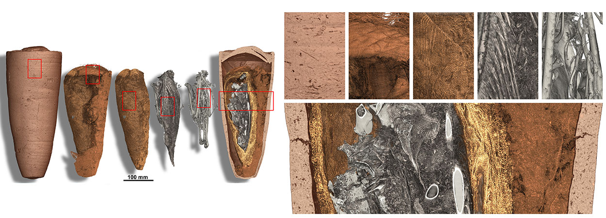
Egyptian Ibis mummy in its sealed jar curated at the Musée de Grenoble. The different
layers are virtually removed: terracotta, loose textile, textile wrapping the animal, soft parts of the
ibis (with feathers) and finally skeleton of the ibis. Credits: Tafforeau, Berruyer, ESRF
A team from the Data Science Research Group (DSRG) at the University of Malta, in collaboration with the European Synchrotron Radiation Facility (ESRF) in France, has recently been awarded a €100,000 grant to research methods for automatically segmenting microtomography images of Egyptian mummies, a process that is currently done manually by trained specialists. The principal investigator on this project is Prof. Johann A. Briffa.
The ESRF synchrotron is a large circular particle accelerator which accelerates electrons to a velocity close to the speed of light. As the electrons pass through certain magnets, they lose energy in the form of synchrotron radiation and X-rays are emitted. These X-rays are then extracted and focused through one of the 44 beamlines coming out of the main accelerator ring. Each beam line is made up of mirrors and specialized optics equipment which guide the X-rays to a sample, allowing scientists to conduct experiments in the fields as diverse as protein biochemistry, earth science, paleontology, materials science, chemistry and physics.
Microtomography is an X-ray imaging technique based on the same principle as the medical CT scanner. Thanks to the extremely powerful X-rays produced in a facility such as ESRF, impressive higher resolution and quality is possible. This can be used to visualize the internal structures of objects in a non-invasive and non-destructive way. Thanks to the phase contrast effect, it can also be used to image soft tissues without the need for contrast agents.
Recently, two of the microtomography beamlines at the ESRF (ID19 and BM05) have been used for applications in Egyptology, through the investigation of animal mummies. In this application, trained specialists manually segment the volume into textiles, organic tissues, balm resin, ceramics and bones. Depending on the complexity and size of the dataset, this process is very time consuming, typically taking several weeks for a small animal mummy. In the near future, thanks to the construction of the new beamline BM18, the same process is expected to be applied to human mummies, which would take considerably longer.
The main objective of this work is to develop and use artificial intelligence techniques to automatically perform this laborious process. Following the principles of “Open Innovation, Open Science, Open to the World”, the developed algorithms, data sets, and results will be made available to the general public.
The project is funded by the ATTRACT programme, itself funded by the European Union’s Horizon 2020 programme.


