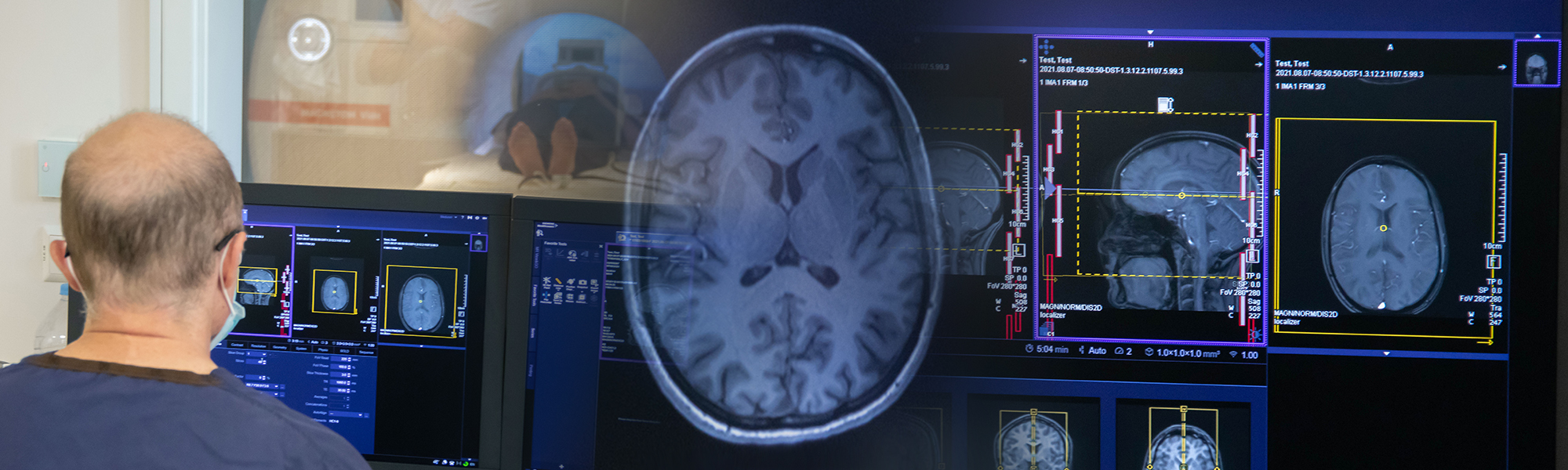
Magnetic Resonance Imaging (MRI) is a powerful, non-invasive technique that uses strong magnetic fields and radio waves to create detailed pictures of the inside of the body. In medicine, MRI helps doctors see organs, tissues, and the brain without the need for surgery or harmful radiation. In research, MRI allows scientists to study how the body works, how diseases develop, and how treatments affect patients over time.
The basic principle comes from physics: when we place the body inside a very strong magnetic field, the tiny magnetic properties of hydrogen atoms in our tissues line up with that field. By sending carefully tuned radio signals, we can make these atoms “resonate” and give off signals that are picked up by the scanner. A computer then turns these signals into images that show the structure of organs and tissues with remarkable clarity.
The idea behind MRI comes from discoveries in physics during the 20th century. Nuclear magnetic resonance (NMR), the scientific foundation of MRI, was first described in the 1940s. In the 1970s, researchers developed the first methods to use NMR to create images of the human body, leading to the medical MRI scanners we know today.

Photo by Siemens Healthineers

Structural MRI produces highly detailed images of the brain and body’s anatomy. It shows the size, shape, and structure of tissues, making it useful for spotting changes caused by injury, disease, or development. Researchers use structural MRI to measure brain volume, detect abnormalities, and track how the brain changes as people grow, age, or respond to therapies.

Functional MRI looks not at structure, but at activity. By measuring changes in blood flow in the brain, fMRI shows which areas are active when a person is thinking, moving, or feeling. This helps researchers understand how different parts of the brain work together and how brain activity changes in conditions such as stroke, dementia, or mental health disorders.
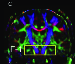
Diffusion MRI focuses on how water moves through tissues, especially in the brain. Because water flows along nerve fibers, this technique can map out the brain’s wiring and connections. Researchers use diffusion MRI to study brain networks, track damage from injury, and better understand diseases that affect the brain’s communication pathways.
MRI Images by M. Pietzuch et al.